
Order Total (1 Item Items):
Shipping Destination:

Showing results for Torsten Bert Moller (219 results)
Still search for
torsten bert mller
Skip to main search results
Search filters
Product Type
- All Product Types
- Books (219)
- Magazines & Periodicals (No further results match this refinement)
- Comics (No further results match this refinement)
- Sheet Music (No further results match this refinement)
- Art, Prints & Posters (No further results match this refinement)
- Photographs (No further results match this refinement)
- Maps (No further results match this refinement)
- Manuscripts & Paper Collectibles (No further results match this refinement)
Condition Learn more
Collectible Attributes
- First Edition (No further results match this refinement)
- Signed (No further results match this refinement)
- Dust Jacket (1)
- Seller-Supplied Images (137)
- Not Print on Demand (219)
Language (3)
Free Shipping
Seller Location
Seller Rating
-
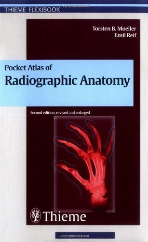
Pocket Atlas of Radiographic Anatomy
Language: English
Published by Thieme Publishing Group, Germany, Stuttgart, 1999
ISBN 10: 3137842026 ISBN 13: 9783137842026
Seller: WorldofBooks, Goring-By-Sea, WS, United Kingdom
Seller rating 5 out of 5 stars

Paperback. Condition: Very Good. This work provides a quick guide to identifying anatomic structures in all commonly performed conventional radiographic examinations. In this edition, many studies have been replaced with better quality radiographs and drawings. The book has been read, but is in excellent condition. Pages are intact and not marred by notes or highlighting. The spine remains undamaged.
-

Pocket Atlas of Radiographic Positioning (Thieme flexibooks)
Language: English
Published by Thieme Publishing Group, 1996
ISBN 10: 3131074418 ISBN 13: 9783131074416
Condition: Very Good. Most items will be dispatched the same or the next working day. A copy that has been read, but is in excellent condition. Pages are intact and not marred by notes or highlighting. The spine remains undamaged.
-

Pocket Atlas of Radiographic Positioning (Thieme flexibooks)
Language: English
Published by Thieme Publishing Group, 1996
ISBN 10: 3131074418 ISBN 13: 9783131074416
Condition: Good. Most items will be dispatched the same or the next working day. A copy that has been read but remains in clean condition. All of the pages are intact and the cover is intact and the spine may show signs of wear. The book may have minor markings which are not specifically mentioned.
-

Paperback. Condition: Good. 1. It's a preowned item in good condition and includes all the pages. It may have some general signs of wear and tear, such as markings, highlighting, slight damage to the cover, minimal wear to the binding, etc., but they will not affect the overall reading experience.
-

Pocket Atlas of Radiographic Anatomy (Flexibooks)
Seller: Goodwill Southern California, Los Angeles, CA, U.S.A.
Seller rating 5 out of 5 stars
Condition: acceptable.
-

Pocket Atlas of Radiographic Anatomy (Flexibooks)
Seller: Goodwill Southern California, Los Angeles, CA, U.S.A.
Seller rating 5 out of 5 stars
Condition: like_new.
-
Pocket Atlas of Sectional Anatomy, Volume III: Spine, Extremities, Joints: Computed Tomography and Magnetic Resonance Imaging
Seller: World of Books (was SecondSale), Montgomery, IL, U.S.A.
Seller rating 5 out of 5 stars
Condition: Very Good. Item in very good condition! Textbooks may not include supplemental items i.e. CDs, access codes etc.
-

Condition: acceptable. This book is in acceptable condition and may have highlighting and or writing throughout. The actual cover image may not match the stock photo, dust jacket may be damaged or missing. Book may show internal and or external wear on spine or cover and may be slightly skewed or have creased pages. This is a used book so codes may be invalid or accompanying media may be missing. May be an Ex library book with stickers and stamps.
-
Condition: Gebraucht - Gut. 1997 - Thieme Verlag - h4.
-

Condition: good. Fast Free Shipping �" Good condition book with a firm cover and clean, readable pages. Shows normal use, including some light wear or limited notes highlighting, yet remains a dependable copy overall. Supplemental items like CDs or access codes may not be included.
-

Condition: good. Fast Free Shipping �" Good condition book with a firm cover and clean, readable pages. Shows normal use, including some light wear or limited notes highlighting, yet remains a dependable copy overall. Supplemental items like CDs or access codes may not be included.
-
-

Pocket Atlas of Sectional Anatomy, Volume I: Head and Neck
Language: English
Published by Thieme Publishing Group, DE, 2013
ISBN 10: 3131255048 ISBN 13: 9783131255044
Seller: Rarewaves.com USA, London, LONDO, United Kingdom
Seller rating 5 out of 5 stars
Paperback. Condition: New. Fourth Edition. This comprehensive, easy-to-consult pocket atlas is renowned for its superb illustrations and ability to depict sectional anatomy in every plane. Together with its two companion volumes, it provides a highly specialized navigational tool for all clinicians who need to master radiologic anatomy and accurately interpret CT and MR images. Special features of Pocket Atlas of Sectional Anatomy:Didactic organization in two-page units, with high-quality radiographs on one side and brilliant, full-color diagrams on the other Hundreds of high-resolution CT and MR images made with the latest generation of scanners (e.g., 3T MRI, 64-slice CT) Consistent color coding, making it easy to identify similar structures across several slices Concise, easy-to-read labeling of all figuresUpdates for the 4th edition of Volume I: New cranial CT imaging sequences of the axial and coronal temporal bone Expanded MR section, with all new 3T MR images of the temporal lobe and hippocampus, basilar artery, cranial nerves, cavernous sinus, and more New arterial MR angiography sequences of the neck and additional larynx images Compact, easy-to-use, highly visual, and designed for quick recall, this book is ideal for use in both the clinical and study settings.
-
Pocket Atlas of Sectional Anatomy: Spine, Extremities, Joints Volume 3: Computed Tomography and Magnetic Resonance Imaging
Language: English
Published by Thieme Medical Publishers 2017-02-10, 2017
ISBN 10: 3131431725 ISBN 13: 9783131431721
US$ 58.09
US$ 20.72 shipping
Ships from United Kingdom to U.S.A.Quantity: 3 available
Add to basketPaperback. Condition: New.
-

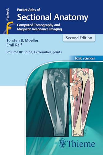
Pocket Atlas of Sectional Anatomy, Volume III: Spine, Extremities, Joints
Language: English
Published by Thieme Publishing Group, DE, 2016
ISBN 10: 3131431725 ISBN 13: 9783131431721
Seller: Rarewaves.com USA, London, LONDO, United Kingdom
Seller rating 5 out of 5 stars
Paperback. Condition: New. Second Edition. Full multiplanar coverage of the spine, extremities, and joints! Renowned for its superb illustrations and highly practical information, the third volume of this classic reference reflects the very latest in state-of-the-art imaging technology. Together with Volumes 1 and 2, this compact and portable book provides a highly specialized navigational tool for clinicians seeking to master the ability to recognize anatomical structures and accurately interpret CT and MR images. Highlights of Volume 3: New CT and MR images of the highest qualityDidactic organization using two-page units, with radiographs on one page and full-color illustrations on the nextConcise, easy-to-read labeling on all figuresColor-coded, schematic diagrams that indicate the level of each sectionSectional enlargements for detailed classification of the anatomical structureComprehensive, compact, and portable, this popular book is ideal for use in both the classroom and clinical setting.
-
Pocket Atlas of Sectional Anatomy, Vol. 1: Head and Neck, Computed Tomography and Magnetic Resonance Imaging, 4th Edition
Seller: Ria Christie Collections, Uxbridge, United Kingdom
Seller rating 5 out of 5 stars
US$ 66.65
US$ 16.03 shipping
Ships from United Kingdom to U.S.A.Quantity: 2 available
Add to basketCondition: New. In.
-

Condition: As New. Unread book in perfect condition.
-
Paperback. Condition: New. In shrink wrap. Looks like an interesting title!
-
Condition: New.
-
Condition: New.
-

Pocket Atlas of Sectional Anatomy, Vol. 2: Thorax, Heart, Abdomen and Pelvis
Language: English
Published by Georg Thieme Verlag Sep 2013, 2013
ISBN 10: 3131256044 ISBN 13: 9783131256041
Seller: Rheinberg-Buch Andreas Meier eK, Bergisch Gladbach, Germany
Seller rating 5 out of 5 stars
Taschenbuch. Condition: Neu. Neuware -This comprehensive, easy-to-consult pocket atlas is renowned for its superb illustrations and ability to depict sectional anatomy in every plane. Together with its two companion volumes, it provides a highly specialized navigational tool for all clinicians who need to master radiologic anatomy and accurately interpret CT and MR images.Special features of Pocket Atlas of Sectional Anatomy:- Didactic organization in two-page units, with high-quality radiographs on one side and brilliant, full-color diagrams on the other- Hundreds of high-resolution CT and MR images made with the latest generation of scanners (e.g., 3T MRI, 64-slice CT)- Color-coded schematic drawings that indicate the level of each section- Consistent color coding, making it easy to identify similar structures across several slicesUpdates for the 4th edition of Volume II:- CT imaging of the chest and abdomen in all 3 planes: axial, sagittal, and coronal- New back-cover foldout featuring pulmonary and hepatic segments and lymph node stations- Follows standard international classifications of the American Heart Association for cardiac vessels and the AJCC/UICC for mediastinal lymph nodes Compact, easy-to-use, highly visual, and designed for quick recall, this book is ideal for use in both the clinical andstudy settings. 328 pp. Englisch.
-

Pocket Atlas of Sectional Anatomy, Vol. 2: Thorax, Heart, Abdomen and Pelvis
Language: English
Published by Georg Thieme Verlag Sep 2013, 2013
ISBN 10: 3131256044 ISBN 13: 9783131256041
Seller: BuchWeltWeit Ludwig Meier e.K., Bergisch Gladbach, Germany
Seller rating 5 out of 5 stars
Taschenbuch. Condition: Neu. Neuware -This comprehensive, easy-to-consult pocket atlas is renowned for its superb illustrations and ability to depict sectional anatomy in every plane. Together with its two companion volumes, it provides a highly specialized navigational tool for all clinicians who need to master radiologic anatomy and accurately interpret CT and MR images.Special features of Pocket Atlas of Sectional Anatomy:- Didactic organization in two-page units, with high-quality radiographs on one side and brilliant, full-color diagrams on the other- Hundreds of high-resolution CT and MR images made with the latest generation of scanners (e.g., 3T MRI, 64-slice CT)- Color-coded schematic drawings that indicate the level of each section- Consistent color coding, making it easy to identify similar structures across several slicesUpdates for the 4th edition of Volume II:- CT imaging of the chest and abdomen in all 3 planes: axial, sagittal, and coronal- New back-cover foldout featuring pulmonary and hepatic segments and lymph node stations- Follows standard international classifications of the American Heart Association for cardiac vessels and the AJCC/UICC for mediastinal lymph nodes Compact, easy-to-use, highly visual, and designed for quick recall, this book is ideal for use in both the clinical andstudy settings. 328 pp. Englisch.
-
Pocket Atlas of Sectional Anatomy, Volume III: Spine, Extremities, Joints: Computed Tomography and Magnetic Resonance Imaging
Seller: Ria Christie Collections, Uxbridge, United Kingdom
Seller rating 5 out of 5 stars
US$ 72.26
US$ 16.03 shipping
Ships from United Kingdom to U.S.A.Quantity: 1 available
Add to basketCondition: New. In.
-

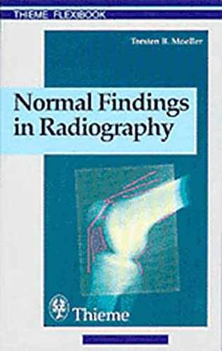
Kartoniert / Broschiert. Condition: New. Torsten B. Moeller, Emil ReifThe key for any beginning radiologist who wishes to recognize pathological findings is to first acquire an ability to distinguish them from normal ones. This outstanding guide gives beginning radiologists the tools they ne.
-

Kartoniert / Broschiert. Condition: New. Torsten B. MoellerThe method of making findings and the systematic method of interpreting images are still relevant today.This book deals with a subject that is seemingly trivial: normal findings in radiology. However,.
-
Pocket Atlas of Sectional Anatomy: Spine, Extremities, Joints Volume 3: Computed Tomography and Magnetic Resonance Imaging
Language: English
Published by Thieme Medical Publishers 2017-02-10, 2017
ISBN 10: 3131431725 ISBN 13: 9783131431721
US$ 67.71
US$ 20.72 shipping
Ships from United Kingdom to U.S.A.Quantity: 1 available
Add to basketPaperback. Condition: New.
-
Condition: New.
-
Condition: New.
-

Pocket Atlas of Sectional Anatomy, Vol. 2: Thorax, Heart, Abdomen and Pelvis
Language: English
Published by Georg Thieme Verlag Sep 2013, 2013
ISBN 10: 3131256044 ISBN 13: 9783131256041
Taschenbuch. Condition: Neu. Neuware -This comprehensive, easy-to-consult pocket atlas is renowned for its superb illustrations and ability to depict sectional anatomy in every plane. Together with its two companion volumes, it provides a highly specialized navigational tool for all clinicians who need to master radiologic anatomy and accurately interpret CT and MR images.Special features of Pocket Atlas of Sectional Anatomy: Didactic organization in two-page units, with high-quality radiographs on one side and brilliant, full-color diagrams on the other- Hundreds of high-resolution CT and MR images made with the latest generation of scanners (e.g., 3T MRI, 64-slice CT)- Color-coded schematic drawings that indicate the level of each section- Consistent color coding, making it easy to identify similar structures across several slicesUpdates for the 4th edition of Volume II: CT imaging of the chest and abdomen in all 3 planes: axial, sagittal, and coronal- New back-cover foldout featuring pulmonary and hepatic segments and lymph node stations- Follows standard international classifications of the American Heart Association for cardiac vessels and the AJCC/UICC for mediastinal lymph nodesCompact, easy-to-use, highly visual, and designed for quick recall, this book is ideal for use in both the clinical and study settings.
-

Pocket Atlas of Sectional Anatomy, Volume III: Spine, Extremities, Joints
Language: English
Published by Georg Thieme Verlag Dez 2016, 2016
ISBN 10: 3131431725 ISBN 13: 9783131431721
Seller: Rheinberg-Buch Andreas Meier eK, Bergisch Gladbach, Germany
Seller rating 5 out of 5 stars
Taschenbuch. Condition: Neu. Neuware -Full multiplanar coverage of the spine, extremities, and joints!Renowned for its superb illustrations and highly practical information, the third volume of this classic reference reflects the very latest in state-of-the-art imaging technology. Together with Volumes 1 and 2, this compact and portable book provides a highly specialized navigational tool for clinicians seeking to master the ability to recognize anatomical structures and accurately interpret CT and MR images.Highlights of Volume 3:- New CT and MR images of the highest quality- Didactic organization using two-page units, with radiographs on one page and full-color illustrations on the next- Concise, easy-to-read labeling on all figures- Color-coded, schematic diagrams that indicate the level of each section- Sectional enlargements for detailed classification of the anatomical structureComprehensive, compact, and portable, this popular book is ideal for use in both the classroom and clinical setting. 480 pp. Englisch.