Items related to Photographic Atlas for Anatomy & Physiology, A

For 2-semester A&P lab course and 1-semester human anatomy lab course
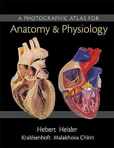
A Photographic Atlas for Anatomy & Physiology is a new visual lab study tool that helps students learn and identify key anatomical structures. Featuring photos from Practice Anatomy Lab ™ 3.1 and other sources, the Atlas includes over 250 cadaver dissection photos, histology photomicrographs, and cat dissection photos plus over 50 photos of anatomical models from leading manufacturers such as 3B Scientific�, SOMSO�, and Denoyer-Geppert Science Company. Two-page spreads with cadaver and anatomical model photos side-by-side help students to better learn and identify structures. The Atlas is composed of 13 chapters, organized by body system, and includes a final chapter with cat dissection photos. In each chapter, students will first explore gross anatomy, as seen on cadavers and anatomical models, and then conclude with relevant histological images.
"synopsis" may belong to another edition of this title.
About the Author:
Nora Hebert, Ph.D. teaches undergraduate courses in Anatomy and Physiology at Red Rocks Community College near Denver, Colorado. Although most of her students are undergraduates, primarily interested in the allied health professions, Nora has also taught graduate-level Human Physiology for the College’s Physician Assistant Program.
Nora is an active faculty member at Red Rocks, serving on the faculty senate, the honors program committee, and the admissions and executive committees for the Physician Assistant Program. She is also part of the College’s Campus Green Initiative.
Among her many academic projects, Nora has consulted in the development of an interactive virtual knee, known as the Explorable Virtual Human, with the Center for Human Simulation at the University of Colorado Health Sciences Center. She has also been involved with the Visible Human Dissector program, advising K-12 teachers and postsecondary instructors on how best to implement the Dissector in their classrooms. Nora has been deeply involved in the development of Practice Anatomy Lab, as coauthor of versions 2.0 and 3.0. She is also the author of over 60 A&P Flix animations covering muscle physiology, neurophysiology, and muscle origins, actions, insertions, and innervations.
Nora received a Ph.D. in Endocrinology from the University of California at Berkeley.
Ruth E. Heisler is a senior instructor in the Department of Integrative Physiology at the University of Colorado at Boulder where she teaches and coordinates several courses, including Human Anatomy, Comparative Vertebrate Anatomy, and Forensic Biology. She has been an instructor at the University of Colorado for more than 15 years.
At the University of Colorado, Ruth has worked extensively with the Science Education Initiative to improve both the teaching and understanding of scientific material at the undergraduate level. In addition, she has been involved in academic outreach through workshops with the American Academy of Forensic Sciences and the Biological Sciences Initiative. She has been a consultant on projects with the Center for Human Simulation, working with data generated through the Visible Human Project.
Ruth has been deeply involved in the development of Practice Anatomy Lab, as coauthor of versions 2.0 and 3.0. She is also author of a custom laboratory manual developed for a large, cadaver-based human anatomy lab.
Ruth received her B.S. in Biology from the University of Minnesota, and her M.A. in Biology from the University of Colorado.
Jett Chinn is an instructor of Human Anatomy in the Science and Technology Division of Ca�ada College (Redwood City, CA) and also the Life and Earth Sciences Department at the College of Marin (Kentfield, CA).
Jett has more than 20 years of experience teaching Human Anatomy at institutions including San Francisco State University, California College of Podiatric Medicine, and Touro University College of Osteopathic Medicine. He has also taught first-year dental students at the UC San Francisco School of Medicine.
Jett received a B.A. in general biology from San Francisco State University.
Karen M. Krabbenhoft, Ph.D. is a senior lecturer in the Department of Neuroscience at the University of Wisconsin in Madison. During her 20-year career, Karen’s focus has been on teaching students at all levels of their educational process, including undergraduate, physician assistant, and medical students. She has been recognized by the University with several awards, including two Medical Alumni Association Distinguished Teaching Awards for the Basic Sciences (1998, 2007), the Dean’s Teaching Award (2000), and the Gender Equity Award (1998). Most recently, Karen was selected by the graduating class to receive the Pre-Clinical Teaching Award in 2011.
Karen earned her Ph.D. in Anatomy from the University of Wisconsin.
Olga Malakhova, M.D. is an assistant scholar in the Department of Anatomy and Cell Biology at the University of Florida College of Medicine in Gainesville. She has been teaching first-, second-, and fourth-year medical students, as well as several Clinical Residency programs, at the University of Florida for the past 20 years.
Olga’s teaching excellence has been recognized by several awards, including six Exemplary Teacher awards from the University of Florida. She was also recognized as a Master Educator by the University’s Medical Education Faculty Development Program (2006).
Olga received her M.D. from Odessa Medical Institute in Ukraine, and her Ph.D. in Neuroscience from the Brain Research Institute of the Russian Academy of Medical Sciences in Moscow.
"About this title" may belong to another edition of this title.
- PublisherPearson
- Publication date2014
- ISBN 10 0321869257
- ISBN 13 9780321869258
- BindingLoose Leaf
- Edition number1
- Number of pages240
- Rating
Buy New
Learn more about this copy
US$ 44.00
Shipping:
US$ 3.99
Within U.S.A.
Top Search Results from the AbeBooks Marketplace
Stock Image
Photographic Atlas for Anatomy & Physiology, A
Seller:
Rating
Book Description loose_leaf. Condition: New. Seller Inventory # mon0000068624
Buy New
US$ 44.00
Convert currency
Stock Image
A Photographic Atlas for Anatomy & Physiology
Seller:
Rating
Book Description Condition: New. loose_leaf. Brand new textbook. Text may have varying degrees of shelf wear and may also have stickers on the covers. Ships fast. Expedited shipping 2-5 business days; standard shipping 7-14 business days. Ships from USA!. Seller Inventory # BUFFALO38-002-967
Buy New
US$ 44.03
Convert currency
Seller Image
Photographic Atlas for Anatomy & Physiology
Seller:
Rating
Book Description Condition: New. Seller Inventory # 20008323-n
Buy New
US$ 67.00
Convert currency
Seller Image
A Photographic Atlas for Anatomy & Physiology
Seller:
Rating
Book Description Condition: New. Seller Inventory # N:9780321869258:ONHAND
Buy New
US$ 65.66
Convert currency
Stock Image
Photographic Atlas for Anatomy & Physiology, A
Seller:
Rating
Book Description Loose Leaf. Condition: New. Brand New!. Seller Inventory # 0321869257
Buy New
US$ 77.21
Convert currency
Stock Image
A Photographic Atlas for Anatomy & Physiology
Published by
Pearson Education 2014-10-24, Boston
(2014)
ISBN 10: 0321869257
ISBN 13: 9780321869258
New
Quantity: 1
Seller:
Rating
Book Description looseleaf in binder. Condition: New. Language: ENG. Seller Inventory # 9780321869258
Buy New
US$ 75.03
Convert currency
Seller Image
Photographic Atlas for Anatomy & Physiology, A (Loose Leaf)
Published by
Pearson Education (US), San Francisco
(2014)
ISBN 10: 0321869257
ISBN 13: 9780321869258
New
Quantity: 1
Seller:
Rating
Book Description Loose Leaf. Condition: new. Loose Leaf. For 2-semester A&P lab course and 1-semester human anatomy lab course A Photographic Atlas for Anatomy & Physiology is a visual lab study tool that helps students learn and identify key anatomical structures. Featuring photos from Practice Anatomy Lab 3.0 and other sources, the Atlas includes over 250 cadaver dissection photos, histology photomicrographs, and cat dissection photos plus over 50 photos of anatomical models from leading manufacturers such as 3B Scientific, SOMSO, and Denoyer-Geppert Science Company. The Atlas is composed of 13 chapters, organised by body system, and includes a final chapter with cat dissection photos. In each chapter, students will first explore gross anatomy, as seen on cadavers and anatomical models, and then conclude with relevant histological images. Shipping may be from multiple locations in the US or from the UK, depending on stock availability. Seller Inventory # 9780321869258
Buy New
US$ 82.40
Convert currency
Stock Image
Photographic Atlas for Anatomy Physiology, A
Seller:
Rating
Book Description Paperback. Condition: new. New. Fast Shipping and good customer service. Seller Inventory # Holz_New_0321869257
Buy New
US$ 79.72
Convert currency
Stock Image
Photographic Atlas for Anatomy Physiology, A
Seller:
Rating
Book Description Paperback. Condition: new. Prompt service guaranteed. Seller Inventory # Clean0321869257
Buy New
US$ 81.40
Convert currency
Stock Image
Photographic Atlas for Anatomy Physiology, A
Seller:
Rating
Book Description UNK. Condition: New. New Book. Shipped from UK. Established seller since 2000. Seller Inventory # IB-9780321869258
Buy New
US$ 86.97
Convert currency